

기술동향
Two heads are better than one
- 등록일2012-11-09
- 조회수5627
- 분류기술동향
-
자료발간일
2012-11-09
-
출처
Riken Research
- 원문링크
-
키워드
#뇌과학#cerebral cortex#human brain#인간 뇌
Two heads are better than one
Comparisons of gene expression in the brains of marmoset monkeys and mice reveal molecular mechanisms underlying evolution of the cerebral cortex
Dramatic expansion of the human cerebral cortex, over the course of evolution, accommodated new areas for specialized cognitive function, including language. Understanding the genetic mechanisms underlying these s, however, remains a challenge to neuroscientists.
A team of researchers in Japan, led by Hideyuki Okano of Keio University School of Medicine and Tomomi Shimogori of the RIKEN Brain Science Institute, has now elucidated the mechanisms of cortical evolution1. They used molecular techniques to compare the gene expression patterns in mouse and monkey brains.
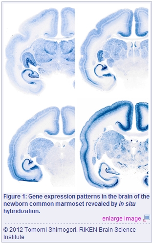
Using the technique called in situ hybridization to visualize the distribution of mRNA trans, Okano, Shimogori and their colleagues examined the expression patterns of genes that are known to regulate development of the mouse brain. They compared these patterns to those of the same genes in the brain of the common marmoset (Fig. 1). They found that most of the genes had similar expression patterns in mice and marmosets, but that some had strikingly different patterns between the two species. Notably, some areas of the visual and prefrontal cortices showed expression patterns that were unique to marmosets.
The researchers observed that the Btbd3 gene, for example, which encodes a tranion factor that regulates the expression of other genes, was expressed throughout the visual cortex of the mouse but restricted to layer 4 of the primary visual cortex, or the V1 area, of the marmoset. Similarly, the gene encoding connective tissue growth factor (CTGF) was expressed throughout the mouse cortex in layer 5, but was restricted to layer 4 of area V1 in the marmoset.
Some of the genes that are expressed widely throughout the mouse prefrontal cortex were likewise restricted to specific layers and sub-regions in the marmoset. Okano, Shimogori and colleagues also noted differences in expression patterns in the subplate region of the developing cortex, which contains the first neurons to receive inputs from the thalamus, a deep brain structure that relays sensory information to the cortex.
The researchers also found differences in gene expression within regions that connect the prefrontal cortex and hippocampus, a structure that is critical for learning and memory.
“The distinct gene expression patterns and anatomical differences between marmosets and mice provide enormous insights into the evolution of the brain,” says Okano. “We are interested in characterizing the functions of genes that could act as driving forces of brain evolution and have ed to investigate several candidate genes. Such approaches will eventually lead to a better understanding of brain function and mental disorders.”
The corresponding author for this highlight is based at the RIKEN?Keio University Joint Research Laboratory, RIKEN Brain Science Institute
- Mashiko, H., Yoshida, A.C., Kikuchi, S.S., Niimi, K., Takahashi, E., Aruga, J., Okano, H. & Shimogori, T. Comparative anatomy of marmoset and mouse cortex from genomic expression. Journal of Neuroscience 32, 5039?5053 (2012). article
-
이전글
- Helping the nervous system pull itself together
-
다음글
- FADD 단백질의 유비퀴틴화 및 분해를 통한 extrinsic apoptosis 및 necroptosis 조절기전
동향